Monday, January 6, 2014

Electrolysis of Water

The Set-Up That I Used:

Materials:

  • Circuit Board
  • 12 volt power adapter
  • Glass Beaker
  • Three Test Tubes
  • 2 68Ω Resistors (For current limiting)
  • Electrical Wire
  • Sodium Bicarbonate
  • Test Tube Clamps and Stand
  • Test Tube Stoppers
  • Tooth Picks


Positive wire: RED
Negative wire: BLUE
Complete set-up


Basic circuit set-up


Procedure:

        First off, I created a mixture of sodium bicarbonate and water. I mixed 200 mL of water and 7 grams of sodium bicarbonate together in an Erlenmeyer flask.
Originally, the test tubes were
completely filled with solution,
but this photo was taken a little
bit after the process started.
        Then, I filled the beaker with 100 mL of the solution. Taking one of the test tubes, I filled it up to the brim with solution, securely stopped it with my finger, inverted it, and lowered it into the beaker until it was submerged. Then I took my finger off and secured it to the test tube clamp. I did the same with a second test tube.
        With the test tubes submerged, I positioned one over the anode and the other over the cathode. Next, I attached the anode and the cathode to the circuit-board, and bubbles started to form on the two wires submerged in the solution.
        I noticed that the hydrogen formed more than two times as fast as the oxygen. I also noticed that the solution turned into a bluish color. These two facts are linked because I think that the blue was copper oxidation from the wires in the solution. For copper oxidation to take place, there needs to be oxygen, a solution, and a current for the copper atoms to combine with the oxygen atoms. That process reduced the amount of oxygen in the water that turned into gas.
Test tube filled with "Hydrogen!"
        When the tube over the cathode was filled with hydrogen, I took a test tube stopper, and with the top of the test tube still underwater, I sealed the test tube, unclamped it, and put it on a test tube rack. I then did the same with the oxygen, but because it was taking so long to fill up, I took it out of the solution when it was only about 1/5 full.
        After I took them out of the solution and sealed them, I took the tube with hydrogen, lit a tooth-pick on fire, unplugged the hydrogen test tube (see video below), and put the flaming tooth pick over the mouth of the test tube. When I put the flaming tooth-pick over the mouth of the test tube, it went "pop!" and the tooth-pick flared up a bit. I did the same thing with the oxygen but instead of a flaming tooth-pick, I blew out the tooth-pick and wile it was still glowing I thrusted it into the  test tube. Unfortunately, because there was not enough oxygen I think, nothing happened. However, when I reopened the oxygen and thrust in a flaming tooth-pick, if you look closely at the video you can see that the flame brightens for a second while it is close to the solution. Finally, I did the same with the mixture of hydrogen and some oxygen that I created by collecting both gases during the electrolysis. It went "POP!" almost immediately after getting in contact with the fire, and the pop it made was louder than the one the hydrogen test tube made. Also, the test tube that contained the hydrogen-oxygen mixture got considerably hot after the gas inside ignited – which did not happen for the test tube containing only hydrogen.


Oxygen test tube

Hydrogen test tube

Mixture of Hydrogen and some Oxygen

Additional Results

I did some additional experiments aimed at studying the effects of the electrolysis process on the metal of the cathode or of the anode. To do so, I used several coins (quarter, nickel) and a wrench. The coins formed very rapidly a green/blue substance when used as the anode, which I think is some oxide of copper. The wrench did not do that, instead, it got kind of corroded when used as the anode. Any of those objects, used as the cathode, got plated with copper. But if they were left in the solution for too long, they acquired a black coating over the copper plating. By the way, the fact that the quarter and the dime both produced a large quantity of green substance means that they are made of copper! In fact, Wikipedia says that quarters and nickels are made up of about 90% copper!


Saturday, June 15, 2013

Color Changes in Winogradsky Columns


      The column that had only paper started out mostly clear, then it started to turn orange, and as time passed the water started to turn dark orange.


      The water in the paper and yolk column also started out mostly clear, turned orange, and then it went more towards a grey color. It also started to grow some algae at the top.


      Like the water in the P column and the P+Y column, the water in the paper, yolk, and shell column started out mostly clear, but then it started to get blacker and darker as time went on. It also started to grow algae on the dirt layer.


      The water in the unenriched column started out dirtier than the other columns, and it got a lot darker as time went on. Either some sort of black plant was living in the column, or a lot of the organisms died, causing the water to darken.


      Going from left to right, the images in the pictures were taken on: Dec 20, 2012, Feb 18, 2013, April 10, 2013, and May 5, 2013.


Sunday, May 5, 2013

Winogradsky Columns Final Day!

Hello! Today I will show you what has happened to the Winogradsky columns. I have extracted a few drops of water from the paper+yolk column, and here are my observations.

P+Y Column Observations

To view the water under my microscope, I used a hanging drop mount. You can see how to make one by reading my previous post "Hanging Drop Mount." 


Figure 2 Small cell with tail
Figure 1 Long
worm-like cell
 In the various water drops I saw some small cells (Fig. 2), long worm-like cells (Fig. 1), and micro-organisms that looked like worms (Fig. 3 & 4). I also saw a cell moving really fast. It could be a paramecium. There were many worm-like bacteria, and a few of the fast moving parameciums. There are also some videos of these organisms below the images.

Figure 4 Two worm like micro-organisms.
about 100µm long

Figure 3 Head of a micro-organism.
about 40µm long


Fast moving paramecium
"Zoo" of organisms. At the top of the image, there
 is a cell that seems to spin when it moves

Video of one of the micro-organisms

Two of the micro-organisms

If you look closely, you can see the long worm
 like cell undulate

Monday, December 10, 2012

Distillation of Ethanol

Ingredients

  • solution of 40% ethanol and 60% water
  • 2 Erlenmeyer flasks
  • hot plate with magnetic stirrer
  • ring stand
  • clamps
  • aluminum foil
  • cotton balls
  • 2 double-hole rubber stoppers
  • thermometer
  • 3 pieces of 4 inch long, 4 mm wide glass tubing
  • 1 piece of 1 1/2 foot silicone tubing
  • 1 glass measuring cup (5 cups)
  • ice 
  • normal water

Procedure

  1. Weigh an empty flask and record the weight for further use.
  2. Pour 200 ml of the ethanol w/ water solution into the flask.
  3. Weigh the flask with the solution.
  4. Subtract the weight of the empty flask from the weight of the flask with the solution. Record for further use. 
  5. Cover the flask with cotton balls then wrap it up in aluminum foil.
  6. Slide one piece of glass tubing into a hole of the stopper, then, in the other hole, slide the thermometer in so it almost touches the bottom of the flask. 
  7. Drop a spin-bar into the flask and close the flask with the stopper that has the tubing and the thermometer in it.
  8. In the other stopper, slide a piece of glass tubing into one hole and another piece into the other hole. One will be your exhaust tube.
  9. Close the second flask with the second stopper.
  10. Put the hotplate to the left of the ring stand. Place the covered flask on top of the hotplate. Do not turn on the hotplate.
  11. Fill the measuring cup with 3 cups water.
  12. Place the measuring cup to the right of the ring stand, and put a clamp onto the ring stand so it hangs over the middle of the measuring cup.
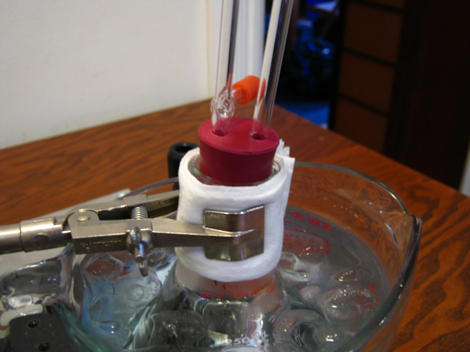
  13. Put the empty flask into the water and clamp it at the neck. Surround the flask with ice.
  14. Attach the silicone tubing to the glass tube on the covered flask, and then attach the other side of the tube to one of the glass tubes on the other flask.
  15. Cover the silicone tubing near the covered flask with more cotton balls and cover it with aluminum foil. This will conserve the heat so the vapor will not condense inside the tubing.
  16. Turn on the hotplate to 120 degrees C and the stirrer to 150 revs./min.
  17. When the thermometer reads about 85 deg. C, the liquid will start to boil. Don't worry, the liquid will stay at that temperature, even with the hot plate a 120 deg. C. The hotplate at 120 deg. C is to make the liquid boil faster and make more vapor.
  18. When the level of liquid in the flask in the water reaches about 30 to 40 ml, turn off the hotplate.
  19. Disassemble every thing so you have one flask with mostly water, and the other flask with probably 90% of ethanol.
  20. Weigh the ethanol in the flask. Subtract the weight of the empty flask from the weight of the flask with ethanol. You will get the weight of the ethanol. Record for further use. 
  21. Store the ethanol for further use and...............
  22. THERES YOUR ETHANOL!
250 ml flask
Stopper with
thermometer


Flask with cotton balls and
aluminum wrap
Setup #1

Setup #2 with silicon tube covered
 with cotton balls and aluminum wrap

Flask in the measuring cup
and ice setup

Glass tube and exhaust pipe setup


My Results

To find my density of the ethanol and of the solution, I divided the weight of the ethanol/solution by the volume in ml of the ethanol/solution. For the density of my solution I got 0.94 g/ml. For the density of the ethanol I got 0.803 ±0.015.


Sunday, November 25, 2012

Calibrating a Microscope

Procedure

  1. Get an iPod with a retina display and place it under a microscope
  2. Using a microscope camera, take a photo of the screen at 10x magnification, using the application ImageJ.
  3. From the documentation, we know that the size of a pixel is 78 µm, corresponding to 326 pixels per inch.
  4. In ImageJ, use the line tool to measure five iPod pixels, and then look at how many pixels of the camera it corresponds to.
  5. Then, go to analyze, and choose set scale.
  6. In set scale, choose the first box and type how many camera pixels the five iPod pixels are.
  7. In the second box type how many µm the five iPod pixels are (390 µm).
  8. Leave the third box alone and in the fourth box write the unit of measurement which is µm.
  9. Then you can also place a scale bar by going to analyze--tools--scale bar and typing how long the scale bar should be, where it should go, and much more.


ImageJ can be found at:
http://rsb.info.nih.gov/ij/download.html

Hanging Drop Mount

Ingredients

  • toothpick
  • pipette
  • coverslip
  • well-slide
  • petroleum jelly
  • drop of stagnant water
  • microscope

Procedure 

  1. Place a coverslip on a clean, flat surface. Use a toothpick to spread a small amount of petroleum jelly around all the edges of the coverslip. Use enough to form a seal between the coverslip and the slide.
  2. Using a pipette, place a drop of the  stagnant water in the center of the coverslip.
  3. Invert a well slide (well side down). Center the well over the coverslip. Use just enough pressure to make sure the coverslip seals to the slide (Don't press to hard or the jelly will ooze into the specimen area).
  4. Invert the assembly so the coverslip is on top. The drop should be suspended in the well from the coverslip).
  5. Observe the slide under a microscope.

Observations

In the drop I found a cluster of organisms. I saw cells, long cells and some moving tiny organisms.


Long cells 100x


Cluster at  45x


More long cells 100x
Four cells 100x


Five different cells 100x


Sunday, November 18, 2012

Gram Staining a Heat Fixed Smear Slide

Ingredients

  • heat fixed smear mount
  • paper towel
  • pipettes
  • distilled water
  • small non corrosive glass or ceramic bowl
  • ethanol
  • lint-free cloth or tissue
  • microscope
  • hucker's crystal violet stain
  • gram's iodine stain
  • safranin O stain

Procedure for Gram Staining

  1. Place a paper towel on a clean, flat surface, then place the heat fixed smear mount onto the towel.
  2. Use a clean pipette to place a drop or two of Hucker's crystal violet stain onto the smear, then use the tip of the pipette (don't touch the smear) to spread the stain until it covers the entire smear.
  3. Allow the Hucker's crystal violet stain to remain in contact with the smear for one minute.
  4. Then, using a pipette filled with distilled water, hold the slide at an angle over the bowl and drip water above the stain so the slide floods with water and washes away any excess stain. 
  5. Drain the slide and place it flat on the paper towel.
  6. Use a clean pipette to place a drop or two of Gram's iodine stain onto the smear. Again, use the tip of the pipette to spread the stain over the entire smear.
  7. Allow the Gram's iodine stain to remain in contact with the smear for one minute.
  8. Fill a clean pipette with ethanol (drugstore 70% ethanol is fine). Hold the slide at an angle over the bowl and drip the ethanol above the smear so it floods the smear. Continue until the ethanol runs colorless.
  9. Then repeat step 4 to rinse all of the ethanol from the slide. Drain the slide and place it flat on the paper towel.
  10. Use a clean pipette to place a drop or two of safranin O stain onto the smear, and again use the tip of the pipette to spread the stain until it covers the entire smear.
  11. Allow the safranin O stain to remain in contact with the smear for one minute.
  12. Repeat step 4 to rinse excess safranin O stain from the slide.
  13. Allow the slide to air dry. If you'r in a hurry, you can gently pat the slide dry with a lint free cloth or tissue. Do not rub the smear area.
  14. Observe the slide under your microscope. You do not need a coverslip.
  15. The Gram-negative bacteria should appear pink or red, and the Gram-positive bacteria should appear violet.


Observations

The bacteria stained only with
Hucker's crystal violet
After staining the smear with Hucker's crystal violet stain, I looked at it under the microscope but the bacteria was to dark to really identify anything except the blobs of bacteria.


The bacteria stained with Hucker's
crystal violet and Gram's iodine
After having stained the smear with Gram's iodine stain, the blobs of bacteria were considerably more transparent. I could also see that some parts of the blobs were lighter in color than the others, which could indicate that those spots were made up of Gram-negative bacteria.


After staining the smear with safranin O, I could clearly see the bacteria at 100x with the different colorations of blue and pink. I also noticed that there is more of the Gram-negative bacteria than the Gram-positive bacteria.


Friday, November 16, 2012

Simple Staining of Epithelial Cells from the Buccal Mucosa

Ingredients

  • normal microscope slide and coverslip
  • microscope
  • toothpick
  • methylene blue stain
  • eosin Y stain
  • paper towel

Procedure for Simple Staining

  1. Transfer one drop of distilled water to the center of a slide.
  2. Use a toothpick to scrape (gently) the inside of your cheek.
  3. Immerse the end of the toothpick in the drop of water and stir to transfer the Epithelial cells to the water.
  4. Position a coverslip over the specimen.
  5. Observe the specimen under a microscope.
  6. Place one drop of methylene blue stain at one edge of the coverslip.
  7. Touch the corner of a paper towel to the slide at the opposite edge of the coverslip. The paper towel wicks the water from under the coverslip, drawing the drop of methylene blue stain under the coverslip.
  8. Allow the stain to work for 30 seconds or so and observe the cells under a microscope.
  9. Repeat steps 6 through 8 with a drop of eosin Y stain instead of methylene blue stain.
  10. Observe under a microscope.

Observations


At 40x with the slide unstained I can see the cells and their nucleus without much difficulty.
At 100x with the slide unstained I can see the nucleus better and also that there are things around it.
At 450x with the slide unstained I clearly see the nucleus and what could be organelles surrounding it.


Epithelial Cells at 40x Non-stained


Epithelial Cells at 100x Non-stained


Epithelial Cell at 450x Non-stained

Due to technical difficulties with the methylene blue stain being to aqueous and made with crystals not fully dissolved, I had the slide all dirty with crystals of methylene blue stain and also the stain did not seem to work. I could not see the cells a lot and could not make any observations on them. Below is a picture of the slide with the bad stain.

The red oval indicates the
 contamination of the
methylene blue stain

Friday, October 19, 2012

Making Sugar Crystals

Making Sugar Crystals

Ingredients and supplies 

  • small saucepan
  • stove
  • wooden stirring spoon
  • sugar
  • 100 ml water
  • pyrex measuring cup (300 ml minimum)
  • wooden skewer 
  • thread

Procedure 


  1. Pour 100 ml of water into the small saucepan and bring to a slight boil.
  2. When the water starts to boil, slowly add sugar while stirring the sugar into the water.
  3. When the sugar starts to accumulate on the bottom of the saucepan immediately transfer the sugar water in to the pyrex measuring cup.
  4. Wrap the thread around the skewer, leaving enough thread hanging of it to almost reach the bottom of the pyrex measuring cup, and secure it.
  5. Slowly submerge the thread into the sugar water and rest the skewer on the brim of the cup.
  6. When the sugar starts to cool, it will form crystals on the thread.
  7. After about a week, crack any crystals on the surface and pull out the thread.
  8. Now you can eat the crystals you made yourself!
The surface crystals
The crystals on the thread
The crystals seen from the side


Friday, October 5, 2012

Winogradsky Colums

Making The Winogradsky Columns

Ingredients

  • 4 tomato sauce jars (about 0.75 liters)
  • 1 gallon of soil
  • 1 egg yolk 
  • 1 egg shell
  • little strips of paper
  • about 1 liter of water

Procedure

  1. In a bowl I mixed enough soil to half fill three jars with the strips of paper.
  2. Then I filled 1 jar half way with the paper enriched soil
  3. After that I mixed the egg yolk into the soil in the bowl.
  4. With which I filled half way another jar with the paper+yolk enriched soil.
  5. Then I crushed the egg shell into the bowl and mixed it together.
  6. And then I filled half way another jar with the paper+yolk+shell enriched soil.
  7. The remaining soil in the bowl I discarded.
  8. With the last jar I filled it 3 cm. from the top with unenriched soil.
  9. With the other three jars I filled them 3 cm. from the top with unenriched soil above the enriched soil.
(abbreviations: paper enriched jar, P. paper+yolk enriched jar, P+Y. paper+yolk+shell enriched jar, P+Y+S. unenriched jar, U)

Observations of the Winogradsky Columns

Day 8


Paper enriched jar

P-enriched jar

The paper enriched jar has amazing signs of life. The organisms I can see are some sort of worm like things that squirm in every direction and poop out dirt that could possibly come from tunnels they are digging. I can see about five of them, and I can see that they are surrounded by little mounds of dirt poop that are shaped like pellets. One of them is bigger than the others and could be the parent.


P+Y-enriched jar

Paper and yolk enriched jar
Interestingly, in this jar there isn't a little worm but there is a web like thing that stretches across the jar 1 cm. from the bottom and has a orange color. If it is an animal that has made it, I have not seen it yet. It could also be possible that the web is an animal its self because it has just fallen onto the soil in a tube shape. If it turns out to be an animal, I will be extremely shocked.


P+Y+S-enriched jar

Paper, yolk, and shell enriched jar
In this jar there is no living thing that I can see, but I hope that in the days to come something will happen.


U-enriched jar

Unenriched jar
In this jar I can see one of those worm things but nothing else. I hope something will happen in the following days in this jar.


Day 12


P-enriched jar


The worms seem to have grown quite a lot. There are still five of them but three of them are really long.

Worm tunnels (a)
Worm tunnels (c)

Worm tunnels (b)

Worm tunnels (d)


P+Y-enriched jar

In this jar everything seems dead. At the top of the soil there is a ring of black soil, and the overall soil is grayish in color. I hope it will revive its self.


Band of black soil


P+Y+S-enriched jar

This jar also has a black band but it is much smaller than the other jar. The soil is also grayish in color. I also hope it will revive its self.

Band of black soil but smaller


U-enriched jar

In this jar there is only one worm but the soil is orangish in color. The soil could be some sort of clay.


Day 17


P-enriched jar

Every thing in here seems dead. Those worms are nowhere to be seen. Also in this jar the black bands of soil have appeared. 


P+Y-enriched jar

Every thing in this jar is still dead and the water is clouded. Probably the only organisms in here (just like the P-enriched jar) are micro-organisms.


P+Y+S-enriched jar

The water in this jar is yellow maybe because a micro-organism that is yellowish in color is thriving here.


U-enriched

In this jar nothing seems different. The little worm is still alive, and the color of the water has not changed.